I must admit that of all the conditions I have treated, heel pain has perhaps fooled me the most. On my behalf, some of the information I was taught was inaccurate.
I initially thought that the pain symptoms, when first arising in the morning or after sitting for a while and then standing, indicated plantar fascia pain. Combine that with direct pressure over the distal medial calcaneus, which reproduces pain, and you have your diagnosis.
It is true that those symptoms most likely indicate a diagnosis of plantar fasciitis, but wait—there is more work that must be done. Many conditions could be causing pain in the heel.
1. Let us start with number one: plantar fasciitis. First, it involves tearing the longitudinal ligament right where it inserts into the medial distal heel bone. Why does this happen? It typically results from an overload of this tissue, which sometimes can cause a reaction in the heel bone, leading to a spur. However, when a spur occurs, it usually indicates that healing is taking place.
Who is more likely to experience plantar fasciitis? It is not as prevalent in the very young and is more likely to occur after the mid-thirties to early forties. Risk factors include having a dropped arch (overpronation) or a lack of dorsiflexion in the ankle, which limits the proper range of motion to allow the shin to translate forward. Individuals with reduced hamstring length have a shocking nine times higher risk, and those with a body mass index above 35% are three times more likely to experience this condition. Anyone can overwhelm their plantar fascia, especially if they increase loading too quickly—such as adding too many miles at once as a runner.
A differential test involves having the patient walk on their heels; if there is no pain but walking on their toes or the ball of the foot reproduces the pain, this is a good indication of plantar fasciitis. Palpating the medial distal heel bone while bringing the great toe back as far as possible can reproduce the same heel pain.
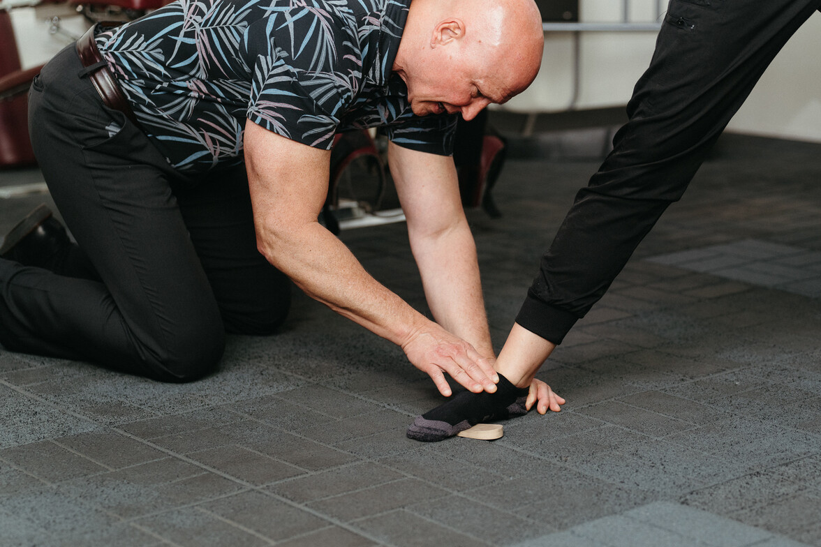
Ways to treat plantar fascia pain include the Strassburg sock, which keeps your foot pulled back while sleeping to create a more functional scar. Soft tissue mobilization—rolling the bottom of the foot, calf, and hamstring—can be beneficial. Stretching exercises for the gastrocnemius, soleus, and hamstring are also recommended. Strengthening exercises like Toe Pro can help. Reducing the amount of time spent on your feet is especially important if you have plantar fasciitis. Heavy compression or shockwave therapy is preferred if there is no inflammation but rather tendinosis.
2. Baxter nerve entrapment involves a branch of the lateral plantar nerve. This can also create heel pain, but the differentiation is that the patient will lose strength in moving the small toe away from the fourth toe, indicating weakness in the muscle abductor digiti minimi. These cases do not respond well to conservative care and typically require referral.
3. Referred pain can also mimic heel pain and is caused by an active trigger point in another muscle that sends pain into the heel. The muscles most likely to replicate heel pain are the abductor hallucis, posterior tibialis, lower medial soleus, medial proximal gastrocnemius, and peroneus tertius. This often becomes a diagnosis where everything else has failed to reproduce symptoms. Diagnosis is made by compressing active trigger points and reproducing the heel pain. Treatment options include active release technique and dry needling overactive trigger points.
4. Neuropathy could involve nerves being stressed within the lumbar spine, canal, or at the intervertebral foramen. If increased pressure in lumbar discs or spinal movement creates symptoms in the heel, that is likely the culprit. Treatment focuses on lowering disc pressure or facilitating the sliding of the nerve root at the foramen.
5. Peripheral neuropathy involves nerve entrapment outside the spinal canal, commonly at the hips' external rotators, hamstrings, and lower leg. Symptoms are often more neurologic and less painful. Treatment includes tissue mobilization using active release or dry needling and nerve flossing, which involves stretching muscles and nerves.
6. I once thought stress fractures of the heel bone, specifically the calcaneus, were very rare, but I was mistaken. The heel bone is designed with a very thin outer layer known as cortical bone to dissipate shock, functioning like a shock absorber. However, this design also makes the bone more prone to fracture. A test for this condition involves placing the medial and lateral heel bones between bilateral hands and applying vice-like compression. The patient may require a walking boot, crutches, or a knee scooter if pain is reproduced. Heel walking is typically very painful, while toe walking should be pain-free.
7. Fat pad syndrome affects about 15% of the population. Patients may experience sharp pain over the center of the heel bone. Diagnosis involves compressing directly over the center of the heel bone to duplicate symptoms. Pain may improve if the fat pad is squeezed inward from medial to lateral and then compressed at the center. Pain tends to worsen when walking on heels and improves with toe walking. For this diagnosis, injections with corticosteroids may be considered, but multiple injections should be avoided, as they can degenerate and cause atrophy of the fat pad, leading to lifelong disability. Effective treatments include heel pads, softer shoes, and avoiding barefoot walking. Taping methods can help position the fat pad more advantageously. Tuli's makes a superior heel cup that we have found effective. This condition also responds favorably to mobilization of the calves, ankle, midfoot, and subtalar joint, as well as improving the strength of the intrinsic foot muscles.
8. Heel pain can also result from tarsal tunnel syndrome. The tarsal tunnel is the area between the heel and the inner ankle. Pain here is often more neurological in sensation (like pins and needles) than sharp. A fallen arch often accompanies it. The best way to evaluate this condition is to compress over the tarsal tunnel at the flexor retinaculum while simultaneously stretching the nerve by dorsiflexing and everting the ankle while flexing and abducting the lower leg and thigh. Reproduction of symptoms during this test strongly indicates the pain's origin. Additionally, compression of the tibial nerve may create weakness in toe flexion. Asymmetrical weakness in toe flexion is another sign of tarsal tunnel syndrome. Symptoms often worsen with prolonged standing, and pain intensifies by day's end. Treatment includes controlling the fallen arch with possible orthotics, soft tissue mobilization, tibial nerve flossing, and specific unloading of the tibial nerve at the flexor retinaculum.
9. Achilles tendinitis or tendinosis will also create heel pain. The Achilles is the strongest tendon in the body and can absorb loads up to twelve times body weight through explosive loading. This will create posterior heel pain. Mobilization and strength of the posterior calf is critical. The best results are achieved with eccentric loading for longer holds. Also, in the short term, a small heel lift can be used on the acute side to reduce the stretch of the Achilles.
This is not meant to be a completely exhaustive list of possible causes of heel pain. However, it has served us well for functional-related heel pain.

.jpg)